For 40 years, the Nikon Small World photomicrography competition has displayed the most beautiful, unusual, and technically sophisticated photographs of a microscopic world that lies just outside normal human experience.
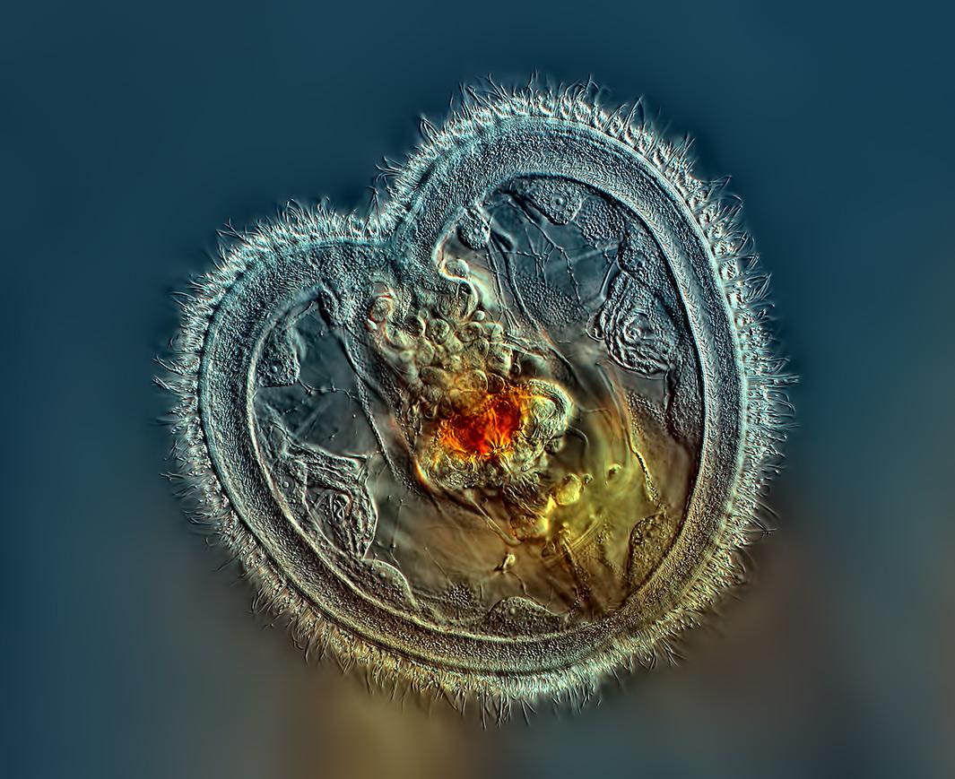
This year’s winning image of a tiny animal called a rotifer is a fascinating example of the beauty of the microscopic world, one full of color, mysterious shapes, textures, and phenomena. The subjects possess a poetry of their own that is at once alien to us but ultimately recognizable.
For Rogelio Moreno, a computer programmer from Panama, taking the winning image and capturing the rotifer in action was a tremendous honor and privilege, a personal accomplishment in a long relationship with science, curiosity, and wonder at the world.
“I’ve always loved science, and since I was little I wanted to understand how things work,” Moreno says. “I read as much as I can, and always wanted to know more.” His first love was astronomy, but the visibility in Panama wasn’t good, since drier, higher elevations are needed for optimal visibility conditions. When Moreno was 12 years old, he got his first microscope for Christmas, which became the main outlet for his curiosity.
Decades later, in the midst of a career as a computer programmer, he became interested in photomicrography after finding photos online. He bought a beginner microscope, but soon that wasn’t enough for all that he wanted to see. He started buying better equipment little by little, reading a lot online and frequenting photomicrography Internet forums to find out about new techniques. There he met people from all over the world—England, Poland, Latin America—who help one another find answers and inspiration. “Anyone who takes this kind of photo knows of the Nikon contest and dreams of winning it, but not as a matter of prestige, but as a platform to show others how fascinating and beautiful the world is.”
The hardest part, Moreno says, as it is with all photography, is to manage all the moving parts that make a good photograph, one that shines, that reaches a place beyond itself. One must have access to the right equipment, the skill and expertise to handle it, and the luck of witnessing the right moment.
“Photography is the science of the moment,” Moreno says, invoking French photographer Henri Cartier-Bresson’s concept of the decisive moment.
And of course, the smaller the subject, the harder it gets to catch these moments. You have to constantly shift your focus. The depth of field becomes narrower and narrower, and after all other factors are accounted for, you have to increasingly rely on luck. In this case, the rotifer shifted just in time, aligning itself to create the suction water current it uses to feed itself and giving a full-on view of its mouth to Moreno.
For years Moreno had been searching for this elusive creature under his lens, especially in a shot that would show the corona around its mouth. The winning rotifer is found in few places in the world, some of them in Moreno’s favorite lake in Panama, Lake Miraflores near Gamboa along the Panama Canal. His wife nicknamed the lake Jurassic Park; the abundant vegetation and animal species located there make for an island ecosystem that can’t be found anywhere else. There he photographed protozoans called ciliates that were found for the first time only in 2007.
Rotifers are a crucial part of the ecosystem that feed on bacteria. Moreno hopes to keep taking photos of these creatures to show others their importance. He will continue to do so by objectively registering the beauty in the world under the microscope.
Below you can see some of the other winning images from this year’s contest.
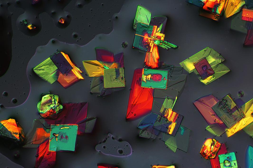
Photo by Chao Zhang/Chinese Academy of Sciences/Nikon Small World
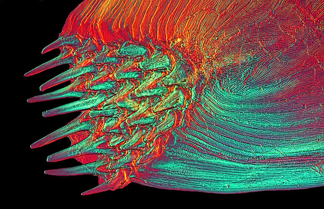
Photo by David Linstead/Nikon Small World
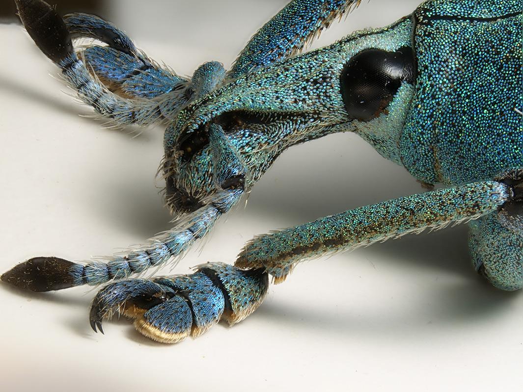
Photo by Luca Toledano/Museo Civico di Storia Naturale di Verona/Nikon Small World
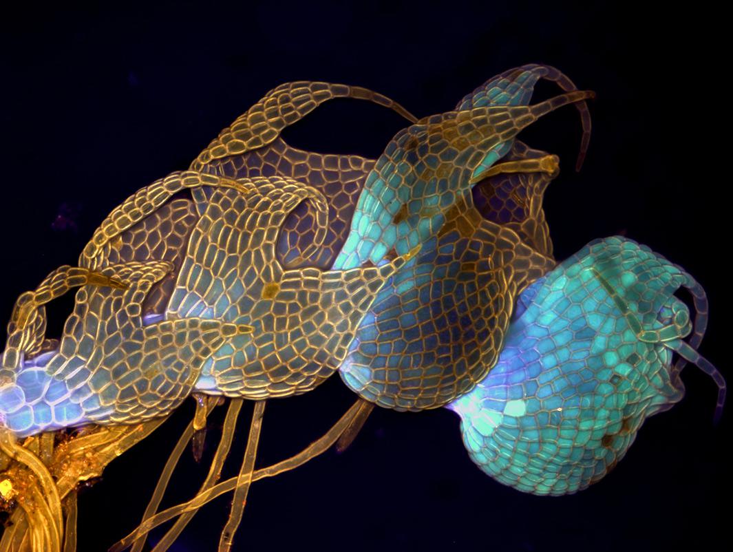
Photo by Magdalena Turzanska/University of Wroclaw/Nikon Small World
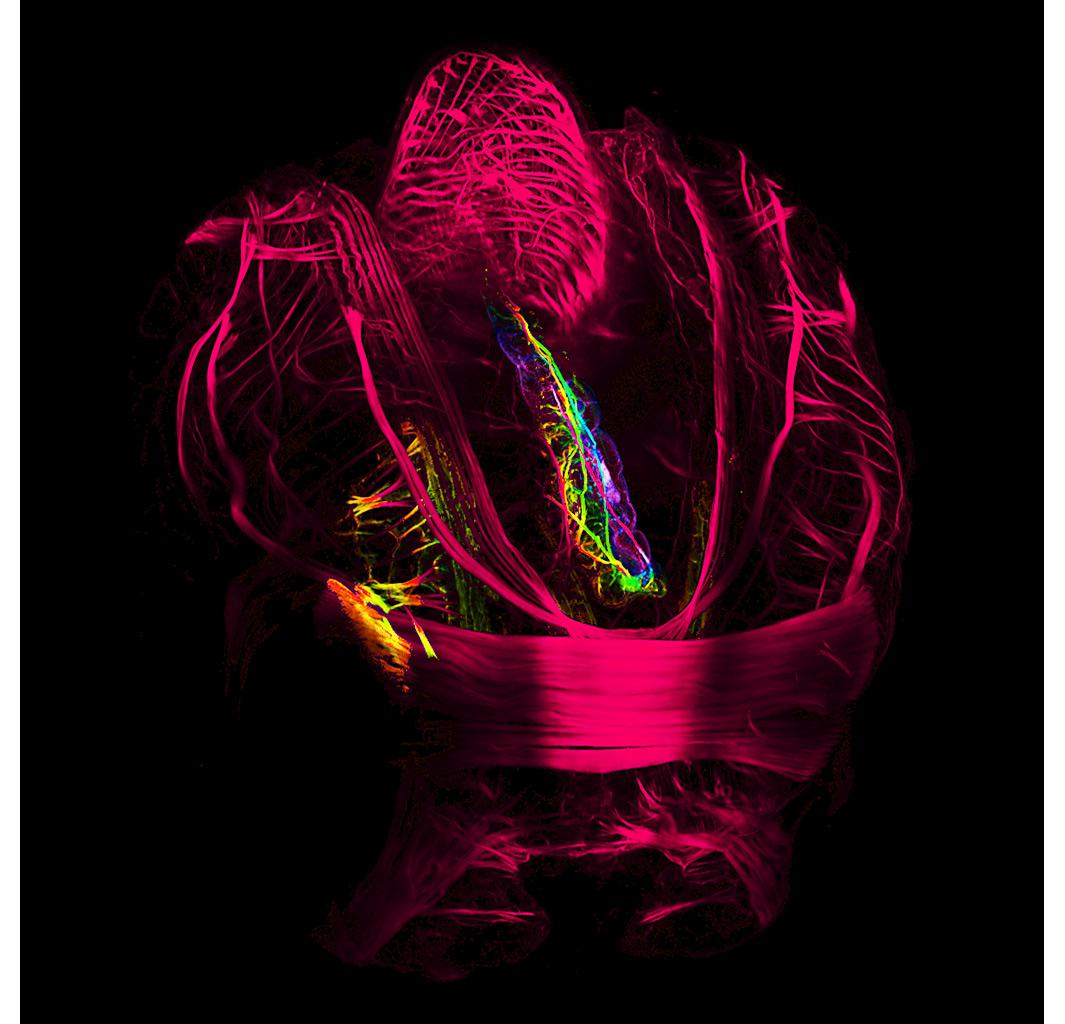
Photo by Andrea Wurzinger-Mayer/University of Vienna/Nikon Small World
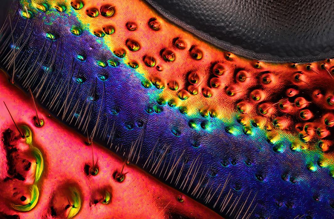
Photo by Charles Krebs Photography/Nikon Small World
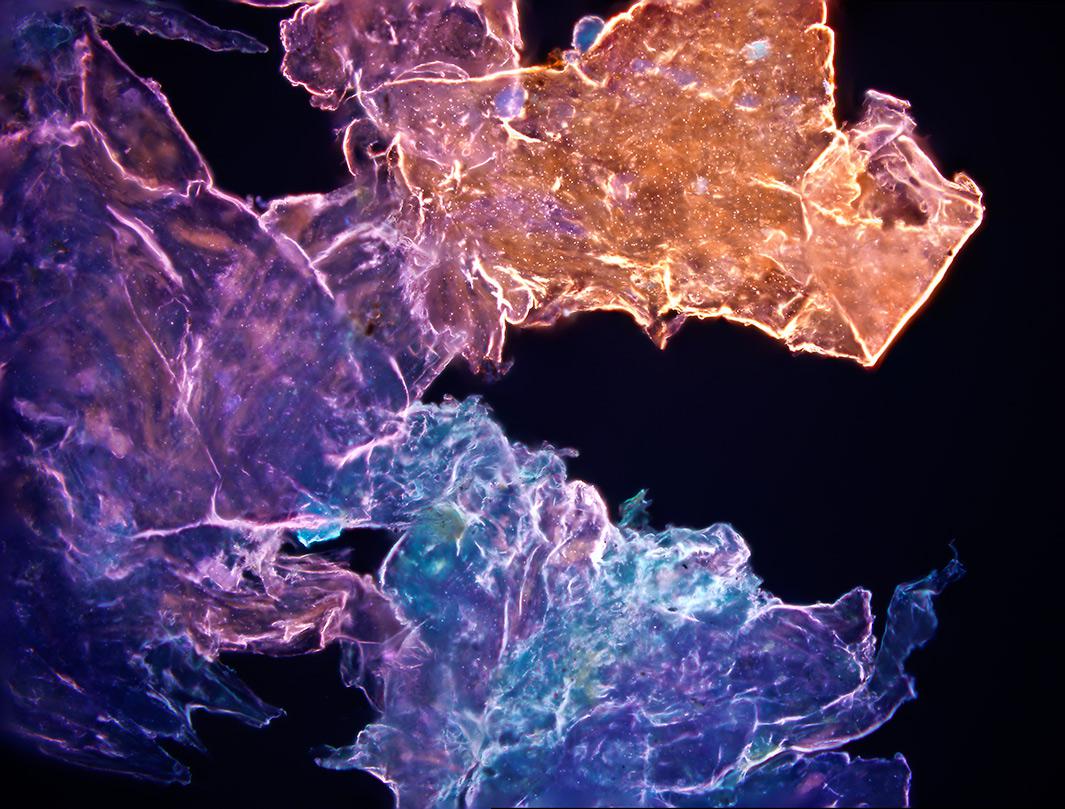
Photo by Daniel Mabel and James Wentzel/Nikon Small World
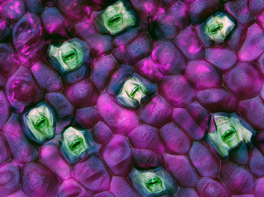
Photo by Dr. Jerzy Gubernator/Faculty of Biotechnology, University of Wroclaw/Nikon Small World
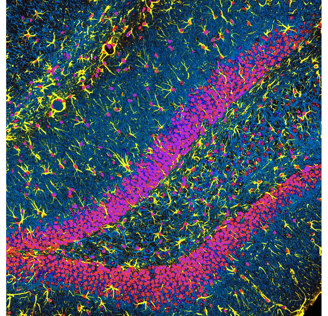
Photo by Dr. Chris Henstridge/MTA-KOKI/Nikon Small World
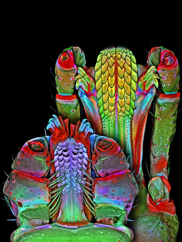
Photo by Dr. Igor Robert Siwanowicz/Howard Hughes Medical Institute/Nikon Small World
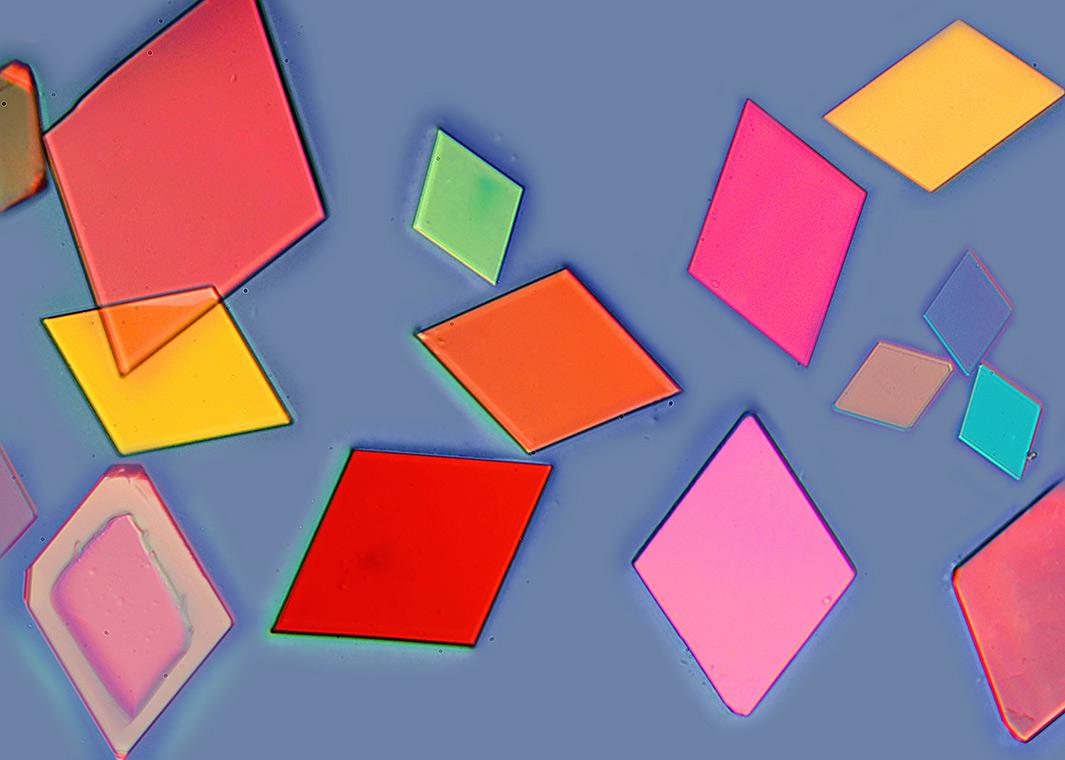
Photo by Yanping Wang/Beijing Planetarium/Nikon Small World